Applications
Application Notes
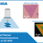
Raman and photoluminescence spectral imaging are applied to reveal otherwise invisible spatial variation in chemical/crystal structure.
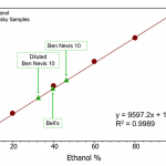
HORIBA ScientificThis application note details how Raman spectroscopy can offer quantitative and qualitative analysis of whisky samples.
Edinburgh Instruments LtdThis note looks at design options to optimise sensitivity, size and wavelength and how they can stretch the limits of applied Raman spectroscopy.
Wasatch PhotonicsHybrid Raman spectroscopy and SERS to detect analytes with low concentrations.
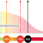
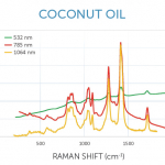
Wasatch PhotonicsUV Raman allows fluorescence background to be eliminated entirely. This is explored in this application note.
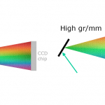
Wasatch PhotonicsThis note describes the considerations in grating selection and how the user may prioritise them.
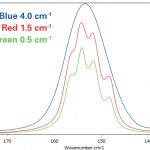
Edinburgh Instruments LtdThis paper discusses how and why the spectral resolution required for dispersive Raman microscopy depends on the intended application and the sample material.
Bruker Optics GmbH & Co. KGThis application note describes how Raman and photoluminescence microscopy can be used as an analysis tool in any forensic laboratory.
Edinburgh Instruments LtdConfocal Raman microscopy is used to study a nicotine patch.
Edinburgh Instruments LtdIn this application note, the use of Raman to monitor the fermentation of glucose (a common feedstock) with yeast is explored.
Wasatch PhotonicsRaman spectroscopy is emerging as a tool to accurately diagnose gout and pseudogout for arthritis sufferers.
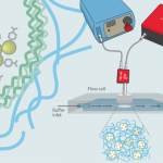
Wasatch PhotonicsSERS has the potential to be use instead of fluorescence-based biosensors. One research group are studying the influence of the supporting hydrogel matrix material on pH assay performance.
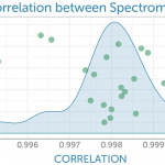
Wasatch PhotonicsA method to correct for slight variations in the wavenumber and intensity response of multiple units is described, achieving better than 99.5 % agreement.
Wasatch PhotonicsPro-Lite describe a new class of spectrometers developed by Wasatch Photonics.
Pro-Lite Technology LtdA RISE system incorporates the advantages of both Raman imaging and SEM in the same vacuum chamber to facilitate the most in-depth characterisation of a sample.
WITec GmbHRaman microscopy is an ideal technique for the analysis of gemstones and other geological samples thanks to its sensitivity to crystalline structures and the presence of minor components within a sample.
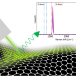
Edinburgh Instruments LtdIn this application note, an Edinburgh Instruments RM5 Raman Microscope is used to highlight how Raman microscopy an essential tool for any material scientist researching graphene.
Edinburgh Instruments LtdPlastic waste is a growing issue and identification of plastic types for separation is key to effective recycling; Raman spectroscopy offers the speed and specificity to help.
Wasatch PhotonicsThis application note describes how Raman microscopy, alone or in combination, can investigate plant cell walls, macrophages and bacteria, and recognise atherosclerosis, differentiate malignant cells and monitor lipid uptake among other capabilities.
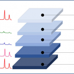
WITec GmbHConfocal Raman imaging is an ideal method for studying 2D materials. It can be used to discern the orientation of their layers and investigate defects, strain and functionalisation.
WITec GmbH

