A Lawrence Livermore National Lab engineer has been awarded $570,000 through the US Department of Energy SunShot Initiative to explore spectroscopic technology as a means of detecting moisture build-up in solar photovoltaic (PV) cells.
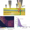
Over the next two years, Mihail Bora, a Materials Engineering Division (MED) research team member at the Lab, will try to prove that spectral imaging can be used to evaluate the moisture content of PV modules and to create two-dimensional maps and models of water concentration. Bora will then use these results as a screening tool to help protect the modules from water damage. Water ingress can cause corrosion of metal parts, delamination and decrease the efficiency of solar cells.
“Our goal is to measure water content without destroying the photovoltaic modules and to remove uncertainty on how reliable the modules are over their long expected lifetimes”, Bora said. “Reliability plays a tremendously important role in making solar competitive with other non-renewable energy sources because it allows project managers to more efficiently use its funds and may lead to a decrease in financing costs.”
Bora’s project builds off the team’s previous testing with laminates and aims to answer questions critical to industry adoption, such as detection range, limits, accuracy and stability. The typical approach to addressing the problem, Bora said, is focused on water transport measurements in polymer membranes or controlled laboratory setups.



![Targeted proton transfer charge reduction (tPTCR) nano-DESI mass spectrometry imaging of liver tissue from orally dosed rat (Animal 3). a) optical image of a blood vessel within liver tissue. b) Composite ion image of charge-reduced haeme-bound α-globin (7+ and 6+ charge states; m/z 2259.9 and m/z 2636.3 respectively, red) and the charged-reduced [FABP+bezafibrate] complex (7+ and 6+ charge states; m/z 2097.5 and m/z 2446.9 respectively, blue). c) Ion image composed from charge-reduced haeme-bound α-globin (7+ and 6+ charge states) showing abundance in blood vessels. d) Ion image composed from charge-reduced [FABP+bezafibrate] complex (7+ and 6+ charge states) showing abundance in bulk tissue and absence in the blood vessel. Reproduced from https://doi.org/10.1002/ange.202202075 under a CC BY licence. Light and mass spectromert imaging of tissue samples](/sites/default/files/styles/thumbnail/public/news/MSI%20drug-protein%20complex-w.jpg?itok=CBNIjyYl)