Police have long relied on fingerprints to identify suspects, however, there has been no way to tell how long ago those prints were left behind: information that could be crucial to a case. A preliminary study in Analytical Chemistry reports that compounds contained in fingerprints could be linked to their age. Scientists have already started mining fingerprint residues for clues to the identity of the person who made them, but timing has proven more difficult to reliably pin down. Notably, past research has shown that a gas chromatography-mass spectrometry method succeeded in determining if prints were more or less than eight days old; however, investigators often need more precision. To get a better idea of when prints were deposited, Young Jin Lee and colleagues looked to reactions already suspected to take place in these residues, when ozone in air reacts with unsaturated triacylglycerols left by a fingertip.
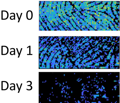
Using prints collected from three donors, the researchers tracked shifting levels of triacylglycerols using mass spectrometry imaging. They found they could reliably determine the triacylglycerol degradation rate for each person over the course of seven days. But the rate differed among individuals, with one person’s triacylglycerols declining more gradually than the others. The researchers attribute this difference to higher levels of lipids in that individual’s fingerprints. The method also worked on residues that had been dusted with forensic powder. The researchers say that although a large-scale study is needed to better understand how lipid levels affect triacylglycerol degradation, this analysis is a first step toward developing a better fingerprint ageing test.


![Targeted proton transfer charge reduction (tPTCR) nano-DESI mass spectrometry imaging of liver tissue from orally dosed rat (Animal 3). a) optical image of a blood vessel within liver tissue. b) Composite ion image of charge-reduced haeme-bound α-globin (7+ and 6+ charge states; m/z 2259.9 and m/z 2636.3 respectively, red) and the charged-reduced [FABP+bezafibrate] complex (7+ and 6+ charge states; m/z 2097.5 and m/z 2446.9 respectively, blue). c) Ion image composed from charge-reduced haeme-bound α-globin (7+ and 6+ charge states) showing abundance in blood vessels. d) Ion image composed from charge-reduced [FABP+bezafibrate] complex (7+ and 6+ charge states) showing abundance in bulk tissue and absence in the blood vessel. Reproduced from https://doi.org/10.1002/ange.202202075 under a CC BY licence. Light and mass spectromert imaging of tissue samples](/sites/default/files/styles/thumbnail/public/news/MSI%20drug-protein%20complex-w.jpg?itok=CBNIjyYl)


