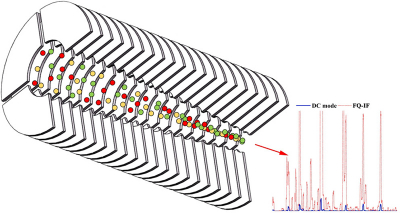
Researchers from the Hefei Institutes of Physical Science (HFIPS) of the Chinese Academy of Sciences (CAS) have proposed a novel focusing quadrupole ion funnel (FQ-IF) that takes advantage of radio frequency ion focusing technology in proton transfer reaction mass spectrometry (PTR-MS) and greatly improves its sensitivity and endowed it with a better “sense of smell”.
PTR-MS is an important analysis technique for the real-time detection of volatile organic compounds (VOCs). Detection sensitivity is a critical performance in PTR-MS. The successful determination of trace VOCs lies on the sensitivity of the instrument, so sensitivity enhancement is an active area of research.
“The FQ-IF drift tube we developed could improve ion transmission efficiency”, said BAO Xun, “and it offers a suitable collision condition”.
The tube consists of 20 layers of stainless-steel electrodes, each with four quarter rings. The first six layers have a constant bore diameter of 22 mm. The later 14 layers taper the inner diameter to 8 mm. The ion transmission efficiency in the drift tube is improved by the radio frequency electric field, which is the key to increase the sensitivity and provides more possibilities for PTR-MS.
The sensitivity of the FQ-IF range now is 13.8–87.9× higher compared to the conventional direct current drift tube, and 1.7–4.8× higher compared to the ion funnel drift tube. The improvements in the limit of detection for the FQ-IF ranged from 2.7× to 35.7× compared to the conventional direct current drift tube. In addition, the FQ-IF drift tube can be easily coupled to other types of mass spectrometers to increase the detection sensitivity and may offer considerable benefits.






![Targeted proton transfer charge reduction (tPTCR) nano-DESI mass spectrometry imaging of liver tissue from orally dosed rat (Animal 3). a) optical image of a blood vessel within liver tissue. b) Composite ion image of charge-reduced haeme-bound α-globin (7+ and 6+ charge states; m/z 2259.9 and m/z 2636.3 respectively, red) and the charged-reduced [FABP+bezafibrate] complex (7+ and 6+ charge states; m/z 2097.5 and m/z 2446.9 respectively, blue). c) Ion image composed from charge-reduced haeme-bound α-globin (7+ and 6+ charge states) showing abundance in blood vessels. d) Ion image composed from charge-reduced [FABP+bezafibrate] complex (7+ and 6+ charge states) showing abundance in bulk tissue and absence in the blood vessel. Reproduced from https://doi.org/10.1002/ange.202202075 under a CC BY licence. Light and mass spectromert imaging of tissue samples](/sites/default/files/styles/thumbnail/public/news/MSI%20drug-protein%20complex-w.jpg?itok=CBNIjyYl)