Tooth whitening is a common aesthetic treatment around the world. To obtain better results, higher concentrations of oxidising agents and longer application times are needed, but this may increase side effects like hypersensitivity and pulp damage, tooth demineralisation and gingival irritation. Besides, the need to apply these products for hours is not very comfortable for the user. Typical tooth whitening treatments are based on the oxidising power of hydrogen peroxide, which breaks the double bonds of the staining molecules on the teeth’s surface making them unable to absorb light. This way the molecule becomes transparent, thus obtaining a bright, clean and white smile.
In recent work from the Research Group of Separation Techniques in Chemistry (GTS) from the Universitat Autònoma de Barcelona (UAB) in collaboration with the ALBA Synchrotron, researchers have used bovine incisors as in vitro model to study the side effects of whitening treatments. They compared typical whitening treatments (based on oxidation with carbamide peroxide) to a new treatment developed and patented by the authors through reduction via metabisulfite, which also makes the staining molecules colourless. However, metabisulfite offers a faster whitening effect, which permits the use of lower concentrations and shorter application times. Results showed how the whitening effect of the novel treatment is highly improved in terms of application time needed, with the consequent reduction of side effects. This makes it a promising candidate to develop novel whitening treatments.
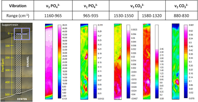
In addition, they found that using oxidising or reducing treatments promotes different mineral changes. This was shown thanks to the chemical imaging performed at the MIRAS beamline of ALBA. It is the first time that synchrotron light is used to map the bovine incisor’s enamel chemically, and to determine the effect of a whitening treatment in terms of chemical mineral modifications, and the extent in deep of these effects.
The team used Fourier transform infrared (FT-IR) microspectroscopy with synchrotron light, which has enabled the combination of biochemical with spatial information at cellular resolution.
“Synchrotron-based Fourier transform infrared microspectroscopy technique, available at the MIRAS beamline in the ALBA Synchrotron, can provide very precise chemical information in small areas, therefore, it is a very suitable technique to study the changes induced by whitening treatments and to characterise the extent of its effects with precision”, explains Ibraheem Yousef, the scientist responsible for the MIRAS beamline.


