Agilent Technologies has announced the opening of a new centre for life science research in partnership with Carleton University in Ottawa, Canada. The Carleton Mass Spectrometry Center, located in the university’s department of chemistry, is equipped with state-of-the-art mass spectrometers, gas and liquid chromatography systems, and bioinformatics tools from Agilent. It will be an analytical resource for researchers and industrial partners across Canada.
“This partnership will enable Agilent to develop new innovative mass spectrometry-based omics workflows for life science research,” said Agilent’s Steve Fischer, marketing director, Academia and Government. “It will make possible new biological discoveries using integrated biology to understand the mechanisms of disease.”
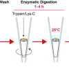
The center is using a new analytical method developed by two of the university’s professors, Jeff Smith and Jeff Manthorpe. The new method, known as TrEnDi (trimethylation enhancement using diazomethane), increases the sensitivity of mass spectrometry analyses by assigning a fixed, permanent positive charge to amino groups. It allows for increased sequence coverage and peptide detection in proteomics analyses, and better detection in metabolomics and lipidomics analyses.


![Targeted proton transfer charge reduction (tPTCR) nano-DESI mass spectrometry imaging of liver tissue from orally dosed rat (Animal 3). a) optical image of a blood vessel within liver tissue. b) Composite ion image of charge-reduced haeme-bound α-globin (7+ and 6+ charge states; m/z 2259.9 and m/z 2636.3 respectively, red) and the charged-reduced [FABP+bezafibrate] complex (7+ and 6+ charge states; m/z 2097.5 and m/z 2446.9 respectively, blue). c) Ion image composed from charge-reduced haeme-bound α-globin (7+ and 6+ charge states) showing abundance in blood vessels. d) Ion image composed from charge-reduced [FABP+bezafibrate] complex (7+ and 6+ charge states) showing abundance in bulk tissue and absence in the blood vessel. Reproduced from https://doi.org/10.1002/ange.202202075 under a CC BY licence. Light and mass spectromert imaging of tissue samples](/sites/default/files/styles/thumbnail/public/news/MSI%20drug-protein%20complex-w.jpg?itok=CBNIjyYl)


